DATABASE AND CLINICAL RECORDS

Our database contains over 18,000 patients’ medical records, which represent the source we draw from every day to daily improve our service. Hospitals, university clinics and health institutions lean on us to access a huge number of files concerning the prosthetic implants of the last 70 years.

Statistical examination from 1985 to 2020 of 18.188 patients registered in our database.

Note: some patients come to our centers many years post-op or trauma with no clinical documentation. In those cases, data are partly reconstructed by deducting them from each patient’s past medical history.

CAUSES LEADING TO ANOPHTHALMOS, EVISCERATION OR SUBATROPHY

(by R. Angeli, University of Milano-Bicocca, 2005)

This study reveals that 53.28% of eye loss cases is determined by accidental events. The most frequent ones happen while playing, as detected in 21.68% of the sample; then, undefined accidents (18.84%), accidents at work (17.52%), home accidents (15.12%), road accidents (11.20%), war-related injuries (7.27%, including WWI, WWII, recent wars and peacekeeping missions), gunshot wounds (4.34%) and unintentional explosions (3.25%, caused by improper use of fireworks, batteries, etc.) are listed.

To sum up, most of the accidents occur at home, very often while playing, or on the workplace. Related traumas not only are contusive and penetrating, but also determined by chemicals like quicklime or solvents. Among chemical burns, the most frequent are the alkali ones – caused by ammonia, caustic soda and lime –, which are even more common than acid burns in domestic and industrial environments. Acids generally and oftentimes involved are the sulfuric, the sulfurous, the acetic, the chloric and the hydrochloric.

Among diseases, neoplasms leading to eye loss represent 14,36% of the reported cases. The most common ones are choroidal melanoma and retinoblastoma, but ocular metastasis, intraocular squamous cell carcinoma, meningioma and lymphoma are diagnosed too.

Among “other diseases“, which concern 23.23% of the sample, glaucoma (30.73%), endobulbar infections or inflammations (24.37%) surgery-related complications like endophthalmitis (14.26%), retinal detachment (13.01%, counting a high relapse rate), and extrabulbar infections like keratitis (11.24%) are listed.

Since the database examined is very large and refers to a long period of time, it is possible to find even 1 case of smallpox – diagnosed in the 1920s – among eradicated diseases. Among other rare pathologies, the following ones are also mentioned: Behcet’s disease, Guillain Barré syndrome, Stringer syndrome, Besser disease, 1 case of typhus and a few ones of ocular TB and syphilis.

Congenital diseases, instead, concern 9.13% of the sample. 42.73% of the related patients suffers from microphthalmos or congenital anophthalmos, 16.60% from congenital glaucoma, 11.58% from congenital cataract, and 29.10% from other congenital diseases, like Von Recklinghausen disease, Goldenhar syndrome, Coats’ disease and Von Hippel-Lindau disease.

(extract of “La gestione del paziente anoftalmico“, by Dr. Raffaella Angeli, University of Milano-Bicocca, supervisor Prof. S. Miglior, assistant supervisor Dr. M. Guareschi.)

CAUSES LEADING TO A SPECIFIC MEDICAL CONDITION

(by R. Angeli, University of Milano-Bicocca, 2005)

Analyzing all factors that may lead to subatrophy, evisceration and enucleation, it is clear that accidental events represent the primary cause behind each of the above-mentioned conditions. As a matter of fact, about 60% of the cases of both evisceration and subatrophy, and about 50% of those of enucleation are determined by accidents.

Among them, the most common ones occur by playing, while being at home or on the workplace. In more detail, play-related accidents are more responsible for subatrophic eyes (25.85%) than enucleated ones (20.68%), while eviscerated eyes are mainly a consequence of work-related injuries.

Because of their metastatic potential, neoplasms appear to be significantly associated with enucleation, and just in a few cases with subatrophy – e.g. after chemotherapy or brachytherapy – or eviscerated eyes.

Other diseases afflict from 20 to 30% of the total number of patients. Among them, retinal detachment mainly leads to subatrophy, while glaucoma, infections and severe endobulbar inflammations mainly cause evisceration and enucleation.

Comparing the above tables, it is evident that some factors are equally responsible for subatrophy, evisceration and enucleation, like gunshot wounds, stab wounds and intentional explosions. Other ones, instead, lead specifically to one of the three conditions, like retinal detachment, that predominantly causes subatrophy (7.54%, compared to 1.97% of enucleations and 2.21% of eviscerations) especially in the event of relapses or after several surgeries.

On the contrary, glaucoma leads to enucleation and evisceration (respectively 7.59% and 9.27%) twice as much as subatrophy (4.86%). That is because enucleation and evisceration surgeries are aimed at killing the pain caused by glaucoma; subatrophic eyes, on the other hand, mostly result from failed surgeries. Therefore, in these cases, subatrophy can be considered as a previous step ending almost unavoidably with enucleation or evisceration.

Eye infections or inflammations, both endobulbar and extrabulbar, lead to subatrophy, evisceration and enucleation respectively as follows: endobulbar ones in 4.71% of the total number of cases and extrabulbar ones in 1.94%, endobulbar ones in 9.13% and extrabulbar ones in 3.60%, endobulbar ones in 5.64% and extrabulbar ones in 2.72%.

Failed surgeries – both directly, because of surgical accidents or complications, and indirectly, because of eye conditions – lead to enucleation in 2.65% of the total number of cases, evisceration in 4.70% and subatrophy in 5.51%. Patients relapsing after retinal detachment were not considered.

COMPARING DALPASSO CLINICAL RECORDS TO EXISTING LITERATURE

The last table compares the clinical records that constitute DALPASSO database to the medical-scientific data found in literature.

Traumatic events


Eye trauma appears to be the predominant cause found in both DALPASSO records (53.28%) and literature (44.28%).

Eye neoplasms


DALPASSO records report that neoplasms determine eye loss in 1,535 patients out of 10,690 (14.36%), that is about half of the percentage averagely registered in literature (33.69%).

Blaydon’s 2003 and Christmas’s 1998 papers underline that neoplasms leading to enucleation are mostly retinoblastoma and choroidal melanoma.

According to Blaydon, retinoblastoma afflicts 1 patient out of 121 (0.8%), causing 30 eviscerations and 91 enucleations; according to Christmas, retinoblastoma afflicts 39 patients out of 342 (11.4%).

As for choroidal melanoma, Blaydon detects 10 cases out of 121 (8.2%) while Christmas reports 104 cases out of 342 (30.4%). In Christmas’s sample, 2 cases of ocular squamous cell carcinoma can be found too.

The huge difference between neoplasms and congenital diseases outlined in the two sources may lie in the kind of centers where these studies were carried out. It is not surprising, indeed, that cancer treatment centers deal with more neoplasms than malformation treatment centers do.

Congenital diseases


In DALPASSO records, congenital diseases afflict 9.13% of the total number of patients, while in literature just 2.59% of the sample. Among all congenital diseases, the most reported ones are undoubtedly microphthalmos and congenital clinical anophthalmos, with a lower presence in literature that cannot be precisely defined since the exact number of cases is unknown. The difference between percentages may be explained by the fact that DALPASSO records reflect the relation of patients to a global population not selected by specific hospital treatments. As an example, congenital glaucoma data can be taken into consideration, since this disease concerns 1.5% of 10,690 DALPASSO cases reflecting likewise the 1:10,000 ratio of patients to population. In Blaydon’s paper, by contrast, congenital glaucoma determines 4.1% of cases corresponding to just 5 out of 121 patients.

Congenital cataract represents 1% of DALPASSO cases, whereas it is not even mentioned in the above-named papers.

Other congenital diseases affect 2.6% of the total number of patients. All pathologies found in literature plus rare cases of Peters’ anomaly and Goldenhar syndrome belong to this group.

In 2003, Blaydon diagnosed 2 cases of fibromatosis (1.6%) and 5 of Retinopathy Of Prematurity (4.1%) out of 121 patients who had lost an eye.

In 1998, during the histopathological analysis of previously enucleated eyes, Christmas diagnosed some cases of Coats’ disease, Persistent Hyperplastic Primary Vitreous and microphthalmos, yet without specifying the number of patients affected.

Other diseases


The most important diseases that belong to this group are retinal detachment, glaucoma, endobulbar infections – like panophthalmitis – and extrabulbar infections – like corneal abscess. This group concerns both sources in a similar way, involving 23.23% of DALPASSO cases and 19.44% of those found in literature.

In literature retinal detachment afflicts 6 patients out of 463 (5.6%), whereas in DALPASSO records it affects 323 patients out of 10,690 (3.02%).

According to Blaydon, end-stage glaucoma, neovascular glaucoma caused by central retinal vein occlusion, and phacolytic glaucoma affect 6.4% (29/463) of patients; according to DALPASSO records, on the other hand, glaucoma in general concerns 7.14% (763/10,690) of the sample.

Ocular infections may lead to eye loss or enucleation too. In literature, 6 cases of endophthalmitis – 3 of which detected by Blaydon in 2003 caused respectively by CMV, toxoplasmosis and toxocariasis – and 3 of corneal ulcer have been diagnosed. As a whole they are 9/463, corresponding to 1.94% of the total number of cases.

In DALPASSO records, on the contrary, extrabulbar infections represent 2.6% of cases (279/10,690) while endobulbar infections affect 5.6% of patients (605/10,690), so infections as a whole concern about 8% of the sample.

Despite the difference between percentages reported in DALPASSO records and in literature is significant, the proportion between endobulbar and extrabulbar infections keeps unchanged, covering respectively 1/3 and 2/3 of total detected infections.

(extract of “La gestione del paziente anoftalmico“, by Dr. Raffaella Angeli, University of Milano-Bicocca, supervisor Prof. S. Miglior, assistant supervisor Dr. M. Guareschi.)

IMPLANTS USE ACCORDING TO A SPECIFIC MEDICAL CONDITION

(by R. Angeli, University of Milano-Bicocca, 2005)

One empty socket and one eviscerated eye constitute the conditions required to consider fitting an implant on a patient.

As this study shows, in the majority of cases the no-implant option is the most adopted one (72.75% of eviscerations and 73.14% of enucleations).

Since the first table outlines over 50 years of researches without taking into consideration the evolution of implants use through time, the no-implant choice made in such a large number of cases may be connected to the absence of a specific rehab procedure for anophthalmic patients. As a matter of fact, in the past, functional rehab was the most important one, and both esthetic and social ones were always omitted.

The second table shows the difference between implants use before and after 1990. Despite the no-implant option yet is the most adopted one, it is important to highlight a trend reversal. Patients with no implants before 1990 were 88.55% on average (85.28% enucleated, 91.82% eviscerated), but after 1990 the percentage lowers to 64.95% (62.25% enucleated, 67.65% eviscerated). This proves that successful treatments for anophthalmic patients have grown significantly, as well as the consideration given to esthetic rehab.

However, choosing not to fit any implant still remains the most popular option among patients, and it may be due to different concomitant factors. One of them certainly is physicians’ lack of interest in treating empty sockets and dealing with the procedures used to partially solve the problem.

It must be reminded, though, that these data come from a database compiled by paramedics – who didn’t personally follow patients’ surgeries and therapies; plus, they were gathered in many different ways, from medical documentation to patients’ reports or physicians’ consultations. If the information obtained from even just one of these means was inaccurate, the final database would be incomplete and in partial disagreement with reality.

Below are all type of implants and their use.

Implants used according to patients’ medical conditions

As for eviscerated eyes, implants with spherical shape are the most widely used because they best adapt to sclerae excavation. As a matter of fact, no recent cases of semi-integrated or new-Allen implants have been detected.

It is interesting to notice that 6 Fox implants can be found in this database. Fox implant, made of a gold sphere, is one of the oldest of all time, and surely left a mark on the history of anophthalmic patients treatment.

As for empty sockets resulting from enucleation, many different types of implants, shapes and materials have been tested, from Allen to the recent Güthoff.

Implants use through the years

The second table shows how implants use has changed through the years according to specific medical conditions – evisceration or enucleation. Before analyzing the data, it is necessary to point out that the total number of cases considered is not 10,690 anymore. What is taken into consideration now is not the patients, but the number of implants fitted. It is possible, indeed, that one patient fits more implants by renewing them in consequence of exposure, extrusion or infection episodes through the years.

The greatest innovation is represented by the introduction of porous implants – hydroxyapatite (HA), Medpor and Güthoff – in the early 1990s. As a matter of fact, before 1990 only 10 HA implants and 1 Medpor were fitted on enucleated patients, in addition to just 1 HA implant on one eviscerated eye.

Since 1990, instead, their use has significantly increased as follows: among enucleated patients, 339 wear HA implants, 25 wear Medpor and 7 wear Güthoff; among eviscerated ones, 55 wear HA and 14 wear Medpor.

At the same time, the overall decrease in non-porous implants use can be noticed. Over the years, non-porous non-spherical implants have suffered a severe reduction – Strampelli from 26 to 2 implants, Bangerter from 69 to 53, new-Allen from 76 to 44 -, whereas spherical ones are by far the most used. Silicone and PMMA implants, indeed, seem to face a trend reversal, passing respectively from 18 to 178 cases (from 0.3% to 5% approximately) and from 282 to 580 (from 5.7% to 13% approximately).

This may be seen as an important step towards the best implant assessment in terms of motility and complications. As a matter of fact, many underline that, having few long-term complications and conferring as much motility as porous implants with no peg do, low cost non-porous implants are the most suitable ones.

As for both non-porous and semi-integrated Allen’s, they still play an important role in this scenario, despite their use has decreased from 6.39% to 6.33% among enucleated patients. Modern surgical techniques, indeed, allow prosthetists to fit implants below Tenon’s capsule, ensuring patients good prosthetic motility, relatively low costs and a percentage of complications comparable to that of non-porous implants.

(extract of “La gestione del paziente anoftalmico“, by Dr. Raffaella Angeli, University of Milano-Bicocca, supervisor Prof. S. Miglior, assistant supervisor Dr. M. Guareschi.)

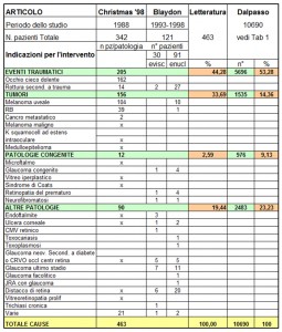
POROUS AND NON-POROUS IMPLANTS

(by R. Angeli, University of Milano-Bicocca, 2005)

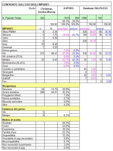
The comparison between DALPASSO records and ASPORS (American Society of Ophthalmic Plastic and Reconstructive Surgery) files highlights some differences.

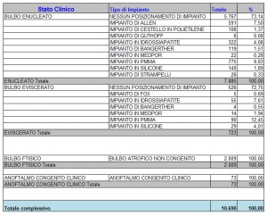
While ASPORS mainly uses porous implants, DALPASSO clearly opts for non-porous ones.

As for porous implants, ASPORS uses Medpor and hydroxyapatite respectively in 42.5% and 26.6% of both evisceration and enucleation cases; on the contrary, DALPASSO opts for hydroxyapatite in 24.8% of enucleation cases and in 33.3% of evisceration ones, and for Medpor in just 1.8% of enucleation cases and in 8.5% of evisceration ones.

As for non-porous implants, DALPASSO works with silicone in 10.8% of enucleation cases and in 18.2% of evisceration ones, and with PMMA in 38.1% of enucleation cases and in 36.4% of evisceration ones; on the other hand, ASPORS makes no distinction between silicone and Medpor, so it uses a non-porous material in 19.9% of enucleation cases and in 25.7% of evisceration ones.

Consequently, these data reveal that Italian cases occurred over the last decades were solved mostly by means of non-porous implants.

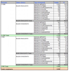
In DALPASSO records, year 1990 represents the cut-off chosen to split time in two periods of analysis. It allows to study the evolution of orbital implants underlining the growing importance of porous ones.

Despite non-porous implants prevail, indeed, the increasing number of porous ones proves that they have definitely turned to be a valid alternative.

This cut-off, however, limits the analysis itself to what happened right before and after 1990, not allowing a more precise examination of the most recent years. This may be one of the reasons why the percentage of porous implants use is lower than the non-porous one.

The treatment of anophthalmic sockets after primary enucleation or evisceration is now directed towards porous materials – like Medpor – without the integration of any covers or pegs. As a matter of fact, porous implants were outlined to be providers of high motility even with no peg integration, and able to reduce the percentage of serious complications such as extrusion or infections. Therefore, numerous literature studies were made to demonstrate advantages and disadvantages of these implants.

On the one hand, it was proved that porous implants with no peg ensure as much motility as non-porous ones do, whereas they provide much more motility after a peg integration – because of that, even two pegs on the same implant have been tested.

On the other hand, many complications were pointed out. Some researches highlight that porous implants percentage of exposure is similar to the highest exposure incidence of Allen implants, PMMA and silicone spheres. Some researchers even reported a significantly higher incidence of complications with HA and PP implants.

Among pros, the no need to cover implants – or the choice to do it by means of autologous tissues – reduces the costs associated with their fitting, intraoperative times and surgical morbidity – since there is no moving from a surgical site to another. It may also simplify vascularization procedures, lower the risk of both infections and a foreign body reaction, and limit the incidence of early complications.

Among cons, a peg integration exponentially increases both incidence of complications and the additional costs related to covering materials, further operations, everything linked to the fiber vascularization of the implant – MRI, scintigraphy and any further related complication –, and a custom-made new prosthesis which needs to be adjusted after the integration.

At the same time, non-porous implants – including semi-integrated ones like Allen – have proved to be cheaper and safer since they have a lower incidence on long-term complications.

Therefore, current debates focus on the actual benefit of fitting porous implants on patients who do not want to rely on pegs.

Nowadays, treatments need to be planned according to all available resources, both owned by patients and provided by treatment centers involved. In this sense, it is more common to see porous implants on young patients, who are very determined to integrate a peg in order to reach a perfect prosthetic motility even though it is more demanding in terms of money, time, and potential further complications.

By contrast, a non-porous implant covered with sclera may be the ideal one for older patients, or for who wants to minimize any further therapeutic action.

(extract of “La gestione del paziente anoftalmico“, by Dr. Raffaella Angeli, University of Milano-Bicocca, supervisor Prof. S. Miglior, assistant supervisor Dr. M. Guareschi.)